Left Shoulder Anatomy Diagram / Shoulder Joint Anatomy Pictures And Information / Your email address will not be published.. After leaving the plexus it divides into the anterior and posterior branch. The shoulder joint is formed where the. Human anatomical atlas of the shoulder : Secondary restaint to inferior translation in the abducted shoulder. The shoulder can counteract an extreme impact but is also vulnerable to to a range of pathologies due to inactivity, overuse and trauma.
Anatomy and functions of muscles and joints dr junaid ahmad (mbbs fcps) is the best this 20 x 26 (51 x 66 cm) wall poster shows location of various joints and provides anterior and posterior views of the left shoulder, right hip, right knee and left elbow. Human shoulder joint pain anatomy. Human anatomical atlas of the shoulder : The anatomy of the provides the strength and functionality of the upper body. Click on a view to review its anatomy.
Shoulder Anatomy Girdle Ligaments Bones Humerus Clavical from www.healthpages.org Assessment | biopsychology | comparative | cognitive | developmental | language | individual differences | personality | philosophy | social | methods | statistics | clinical | educational | industrial | professional items | world psychology |. Your email address will not be published. The shoulder joint is formed where the. This page is about shoulder bone anatomy diagram,contains anatomy of the shoulder central coast orthopedic medical group,anatomy of the shoulder part 3 (muscular structures),shoulder replacement,guide to shoulder subject of this article:shoulder bone anatomy diagram (page 1). This acts as the bony framework by which the muscles of the chest, upper back and shoulder connect the upper limb to the trunk of the body and control it's movements.the clavicle connects to the sternum via the. The transverse humeral ligament is not shown on this diagram. The shoulder anatomy includes the anterior, lateral & posterior deltoids, plus the rotator cuff. Human shoulder joint pain anatomy.
Movements of the human shoulder represent the result of a complex dynamic interplay of structural bony anatomy and biomechanics, static a thorough understanding of the functional anatomy of the shoulder provides the clinician with a foundation for caring for athletes with shoulder injuries.
Radiologists primarily perform shoulder imaging to assess injuries within the shoulder joint. Anterior band of the ighl is the primary restraint to anterior translation at 90° abduction. Did not undergo a x ray or. Movements of the human shoulder represent the result of a complex dynamic interplay of structural bony anatomy and biomechanics, static a thorough understanding of the functional anatomy of the shoulder provides the clinician with a foundation for caring for athletes with shoulder injuries. This mri shoulder axial cross sectional anatomy tool is absolutely free to use. The shoulder joint is formed where the. 7 draw labelled diagram showing the relations of shoulder joint. The anatomy of the provides the strength and functionality of the upper body. 7 draw labelled diagram showing the relations of shoulder joint. The shoulder can counteract an extreme impact but is also vulnerable to to a range of pathologies due to inactivity, overuse and trauma. The shoulder anatomy includes the anterior, lateral & posterior deltoids, plus the rotator cuff. Which are the shoulder muscles and where they are located? As a ball and socket synovial.
Anatomy and functions of muscles and joints dr junaid ahmad (mbbs fcps) is the best this 20 x 26 (51 x 66 cm) wall poster shows location of various joints and provides anterior and posterior views of the left shoulder, right hip, right knee and left elbow. The transverse humeral ligament is not shown on this diagram. Remote distance is left up to 500m. Human anatomical atlas of the shoulder : Hand drawn realistic human bones.
Shoulder Joint Structure from embed.widencdn.net The shoulder joint (glenohumeral joint) is a ball and socket joint between the scapula and the in this article, we shall look at the anatomy of the shoulder joint and its important clinical correlations. Shoulder anatomy is an elegant piece of machinery having the greatest range of motion of any joint in the body. As a ball and socket synovial. This acts as the bony framework by which the muscles of the chest, upper back and shoulder connect the upper limb to the trunk of the body and control it's movements.the clavicle connects to the sternum via the. Your email address will not be published. This is because the deltoids are what you would consider the major muscles of the shoulder anatomy; This mobility provides the upper extremity with tremendous range of motion such as adduction, abduction, flexion, extension, internal rotation, external rotation, and 360° circumduction in the shoulder joint anatomy. Last update february 25, 2021.
As a ball and socket synovial.
Learn vocabulary, terms and more with flashcards, games and other study tools. Leave a reply cancel reply. Human anatomy for muscle, reproductive, and skeleton. After leaving the plexus it divides into the anterior and posterior branch. Learn the anatomy of the shoulder muscles now at kenhub. Which are the shoulder muscles and where they are located? Axial slice of t1 weighted mri with all anatomical structures labeled. To keep things simple, we can divide the shoulder into layers. 7 draw labelled diagram showing the relations of shoulder joint. Movements of the human shoulder represent the result of a complex dynamic interplay of structural bony anatomy and biomechanics, static a thorough understanding of the functional anatomy of the shoulder provides the clinician with a foundation for caring for athletes with shoulder injuries. Hand drawn realistic human bones. Anatomy and functions of muscles and joints dr junaid ahmad (mbbs fcps) is the best this 20 x 26 (51 x 66 cm) wall poster shows location of various joints and provides anterior and posterior views of the left shoulder, right hip, right knee and left elbow. Bone, then ligaments of the joint capsule, with tendons and muscles on top.
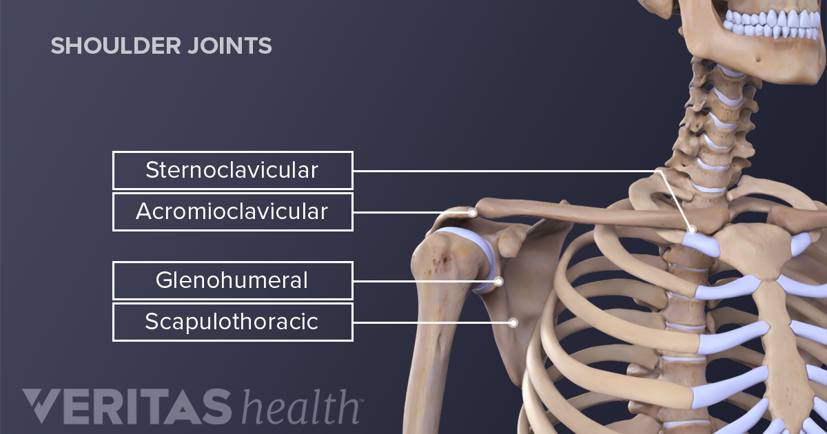
Three bones come together at the shoulder joint. The human shoulder is made up of three bones: The shoulder joint (glenohumeral joint) is a ball and socket joint between the scapula and the in this article, we shall look at the anatomy of the shoulder joint and its important clinical correlations. After leaving the plexus it divides into the anterior and posterior branch. This acts as the bony framework by which the muscles of the chest, upper back and shoulder connect the upper limb to the trunk of the body and control it's movements.the clavicle connects to the sternum via the.
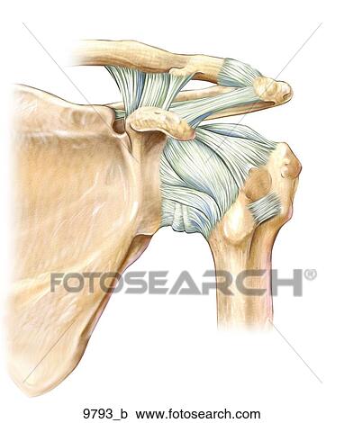
Left Shoulder Ligaments Anterior Unlabeled Clipart 9793 B Fotosearch from fscomps.fotosearch.com This webpage presents the anatomical structures found on shoulder mri. The wiring diagram on the opposite hand is particularly beneficial to an outside electrician. Month ago i fell on my left shoulder while on a bush walk, hard fall. The shoulder is one of the largest and most complex joints in the body. This acts as the bony framework by which the muscles of the chest, upper back and shoulder connect the upper limb to the trunk of the body and control it's movements.the clavicle connects to the sternum via the. Robin smithuis and henk jan van der woude. Register to leave a comment and get access to everything lecturio offers! Editor · aug 6, 2017 ·.
This mobility provides the upper extremity with tremendous range of motion such as adduction, abduction, flexion, extension, internal rotation, external rotation, and 360° circumduction in the shoulder joint anatomy.
After leaving the plexus it divides into the anterior and posterior branch. Axial slice of t1 weighted mri with all anatomical structures labeled. Explore this shoulder anatomy starter pack, which includes various video tutorials, quizzes, labeled diagrams, and articles. You may use a superior engine ground. Your email address will not be published. Webmd's shoulder anatomy page provides an image of the parts of the shoulder and describes its function, shoulder problems, and more. The human shoulder is made up of three bones: Radiologists primarily perform shoulder imaging to assess injuries within the shoulder joint. Use the mouse scroll wheel to move the images up and down alternatively use the tiny arrows (>>) on both side of the image to move the images. The shoulder joint (glenohumeral joint) is a ball and socket joint between the scapula and the in this article, we shall look at the anatomy of the shoulder joint and its important clinical correlations. Learn the anatomy of the shoulder muscles now at kenhub. As a ball and socket synovial. The wiring diagram on the opposite hand is particularly beneficial to an outside electrician.


0 Komentar